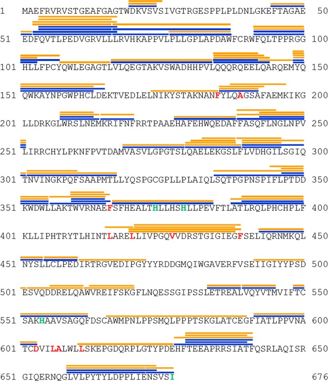
Figure 2.

Peptide map of 15-LOX-2. Peptides were generated by pepsin digest and identified through tandem mass spectrometry. Peptides with measurable ion envelopes are shown as orange lines (in chelated and Ca2+-bound states) and blue lines (in a nanodisc-associated state) above the peptide sequence. Residues that are conserved in mammalian LOXs and thought to be part of substrate binding are colored red, while residues that coordinate the catalytic iron are colored green.
