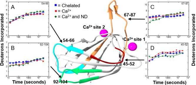
Figure 4.
Impact of Ca2+ binding and membrane association on H/D exchange for 15-LOX-2. The number of deuteriums incorporated on peptides vs time was plotted to obtain H/D exchange kinetics for 15-LOX-2 peptides in a Ca2+ free state (blue squares), a Ca2+-bound state (red circles), and a nanodisc-associated state (green triangles). Peptides are labeled by their primary sequence and mapped onto the PLAT domain of 15-LOX-2 by color: (A) peptide 54–66, (B) peptide 92–104, (C) peptide 67–87, and (D) peptide 45–52.

