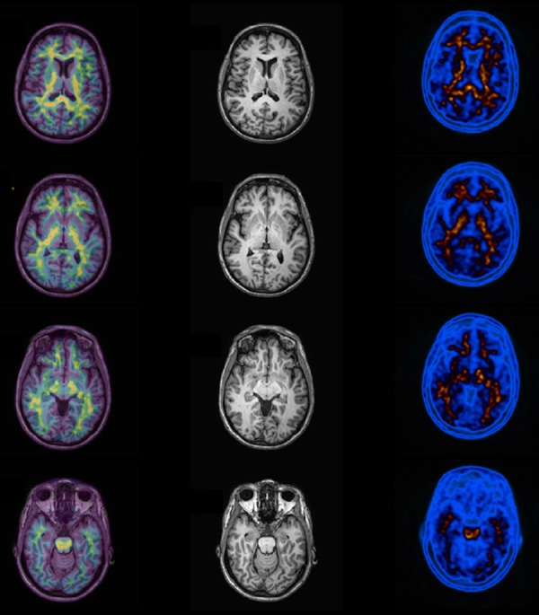
Figure 6.

Normal amyloid PET example. Note normal uptake of PIB (Pittsburg Compound) labeled with carbon-11 in white matter tissue at the left column. In the middle column it is shown the MR images of the patient, and in the right column the fused images (PIB + MRI). No cortical uptake of 11C-PIB is seen in this case.
