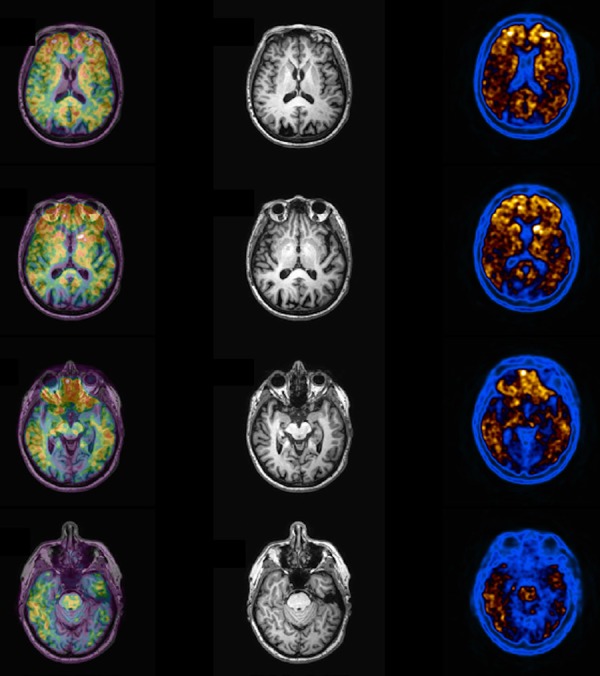
Figure 7.

Abnormal PIB PET imaging a patient with confirmed clinical diagnosis of Alzheimer disease. Note marked uptake in the frontal and parietal cortex, with poor visualization of white matter uptake. Right column is the PIB images, middle the MRI and the left column is seen the fused images (PIB + MRI).
