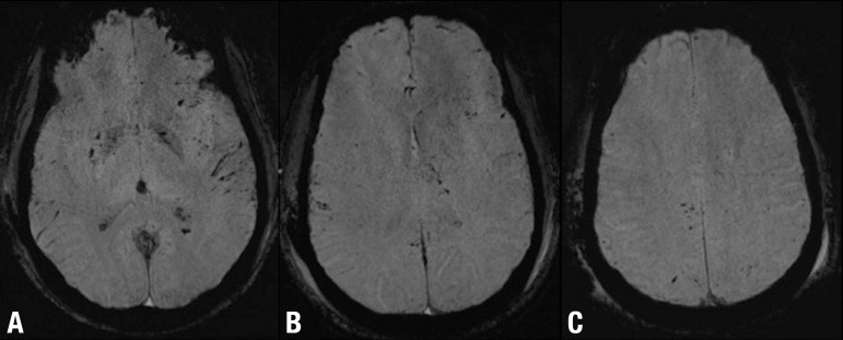
Figure 2.
DAI and subarachnoid hemorrhage after mild TBI. Axial SWI [A, B and C] show multiple focal lesions involving the basal ganglia and the lobar white matter at the gray-white matter interface. Note also the bilateral subarachnoid hemorrhage particularly evident in A B C the left sylvian fissure.

