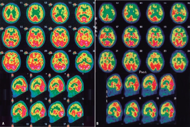
Figure 2.
Brain scintigraphy (SPECT) from patient TA, 48 year-old woman. [A] Brain SPECT after approximately one year since symptoms onset (May/2010): moderate hypoperfusion in medial and dorsolateral prefrontal cortex, with left predominance; very mild hypoperfusion in the left parietal cortex; no hypoperfusion in medial temporal regions. [B] Brain SPECT approximately two years after symptoms onset (March/2011): severe hypoperfusion in prefrontal cortex (with left predominance); severe hypoperfusion in the left parieto-temporal cortex; mild hypoperfusion in the right parieto-temporal cortex; very mild hypoperfusion in the medial temporal regions.

