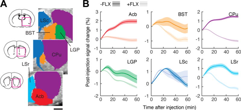
Figure 2.

FLX-dependent MRI time courses in individual brain regions. (A) Definition of six anatomically-based ROIs (colored regions) corresponding to bed nucleus of the stria terminals (BST), caudate-putamen (CPu), lateral globus pallidus (LGP), lateral septum caudal part (LSr) and rostral part (LSc), and nucleus accumbens (Acb). Scale bar = 1 mm. Brain sections (left) show areas of detail (magenta squares) for ROIs centered at the rostrocaudal coordinates indicated in white. (B) Post-injection MRI signal time courses observed in the six ROIs, in animals that did not (dark colors) or did (light colors) receive FLX pretreatment. Shaded margins around each curve denote s.e.m. of six measurements each.
