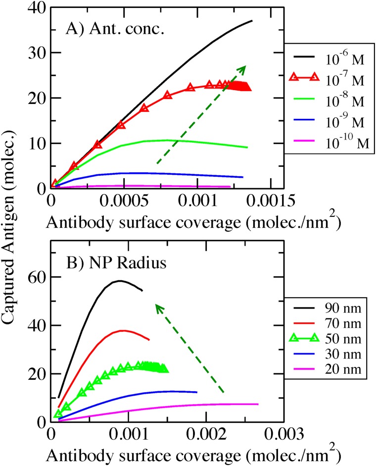
Fig 8. Captured antigen per unit area as a function of antibody surface coverage.
Streptavidin conjugated AcNP, spacer 50 monomers, Kd = 10−9 M. A) For different antigen bulk concentrations. AcNP radius = 50 nm. B) For different AcNP radius. Inset in panel B) are same data but the captured antigen normalized by the area of the NP. Red and black dashed lines, in the inset, represent the ideal situation of all the antibodies bound to two or one antigen respectively. Bulk concentration mix = 100 nM antibody: 200nM antigen. Triangle symbols represent the set of conditions used in this paper for the rest of the calculations.

