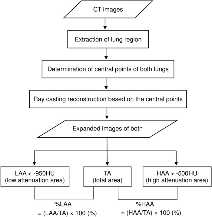
Fig 1. Schematic diagram of 3D-cMPR imaging at a constant depth from the chest wall and assessment.
Overall schematic diagram of the three-dimensional, curved high-resolution CT (3D-cHRCT) image at a constant depth from the chest wall and how it is used for assessment. Total area (TA), low attenuation area (LAA [< -950 HU]), and high attenuation area (%HAA) [> -500 HU]), which were obtained from 3D-cHRCT images, were computed on a workstation using a novel CAD program.

