Figure 5.
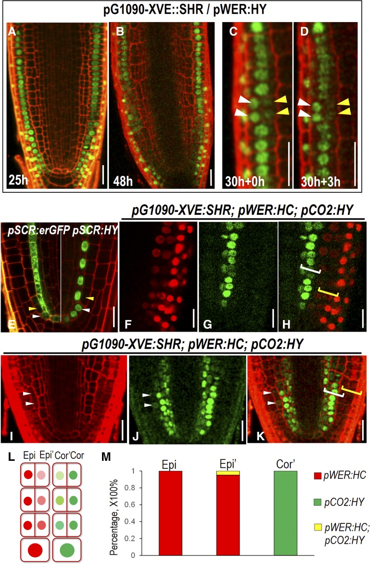
Cell-fate specification in roots with extra cell layers. A and B, Expression of pWER:H2B-YFP (pWER:HY) in pG1090-XVE::SHR roots at different estradiol induction time points. C and D, Time course observation of the same root expressing pG1090-XVE::SHR and pWER:HY at 30 hr estradiol induction (C) and 3 h after (D). White arrowheads point to the expression of pWER:HY in epidermis, and yellow arrowheads point to the expression of pWER:HY in the cell divided from epidermis. E, Expression of pSCR:erGFP (left) and pSCR:HY (right) in wild-type roots. White arrowheads point to CEI and CEID cells in which pSCR is still active. Yellow arrowheads point to the first cortex cell derived from the CEID in which pSCR activity is not seen. F to K, Expression of pWER:HC and pCO2:HY in pG1090-XVE:SHR-expressing roots after 48 h in estradiol. Note the separated expression zone of pWER:HC and pCO2:HY, marked by brackets in H and occasionally overlapped expression zone marked by white arrowheads in I to K. White brackets indicate the expression of pCO2:HY, and yellow brackets indicate the expression of pWER:HC. L, Scheme describing the separated expression zone shown in F to H. Epi, Epidermis; Epi’, extra cell layer derived from epidermis; Cor, cortex; Cor’, extra cell layer derived from cortex. M, Quantification of the percentage of cells expressing different markers in pG1090-XVE:SHR-expressing roots after 48 h in estradiol. Scale bars = 20 μm.

