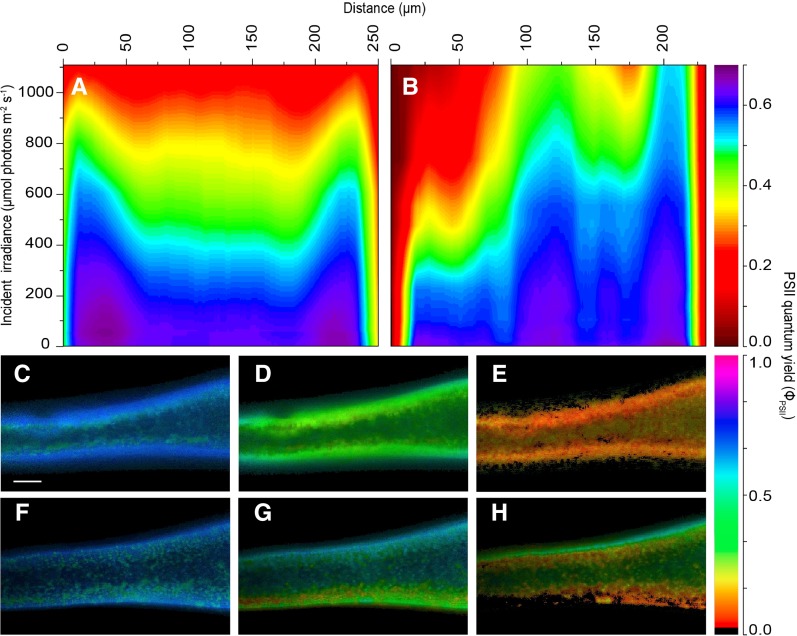
Figure 6.
Isopleths (A and B) and images (C–H) of effective PSII quantum yield in apical thallus fragments of F. vesiculosus illuminated evenly on a cross section or perpendicular on one side of the thallus surface. Images were acquired under red light using either direct light from the built-in LEDs of the variable chlorophyll fluorescence imaging system (590–650 nm) or light perpendicular to the thallus surface provided by a supercontinuum laser (615–665 nm) connected to a tunable single-line filter and delivered via a laser sheet generator. The isopleths (A and B) show the influence of actinic irradiance on the effective PSII quantum yield (in µmol electrons m−2 s−1 [µmol photons m−2 s−1]−1) as a function of the depth in the tissue when illuminated either directly on the cross section (A) or perpendicular to the surface of the thallus (B). Illumination in B was given from left to right. Line profiles (line width = 15 pixels) were taken on thallus parts with similar thickness (∼250 µm), with cortex layers also displaying similar thicknesses (∼50–75 µm). C to H show images of effective PSII quantum yield in darkness, moderate irradiance (567 ± 18 µmol photons m−2 s−1), and saturating irradiance (1,087 ± 30 µmol photons m−2 s−1) under direct even illumination of the cross section (C–E) and with laser light sheet illumination perpendicular to the thallus surface (F–H). Illumination in F to H was given from the bottom to the top. Bar = 0.2 mm.

