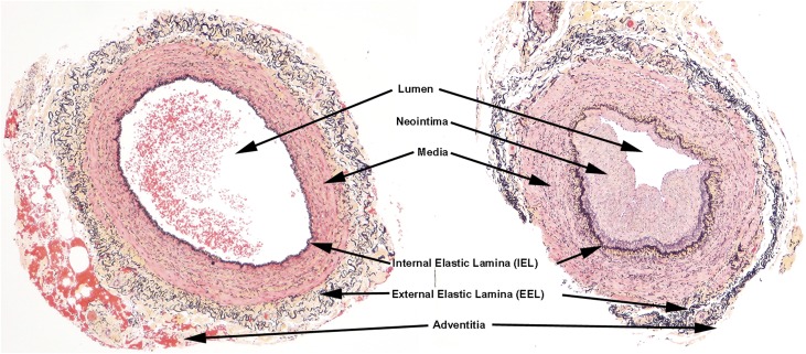
Figure 1.
Illustration of characteristic landmarks of both normal veins and abnormal veins. There is a neointima and accumulation of medial collagen (yellow staining) adjacent to the IEL in the abnormal vein (right). In the abnormal veins, the indicated landmarks were measured morphometrically to determine the extent of luminal compromise by neointima (Movat pentachrome stain).

