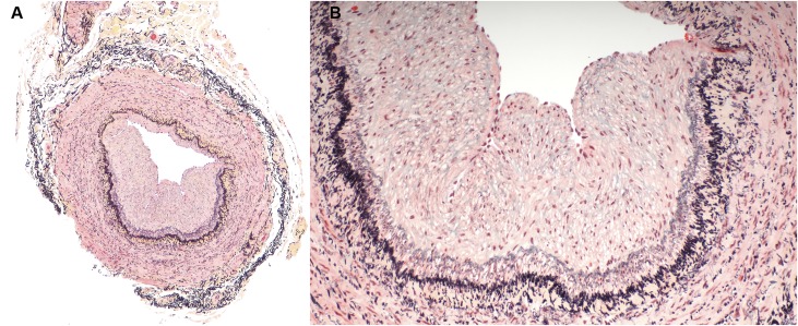
Figure 3.
Abnormal vein with eccentric neointima formation, stained with Movat pentachrome. (A) is a low power image and (B) is a higher power image of the vein section shown on the right in Figure 1. Less proteoglycans (blue staining) are noted in the neointima of this sample and less collagen (yellow staining) is noted in the vessel wall. Inflammatory infiltrates are not present in any of the layers of the vessel wall.

