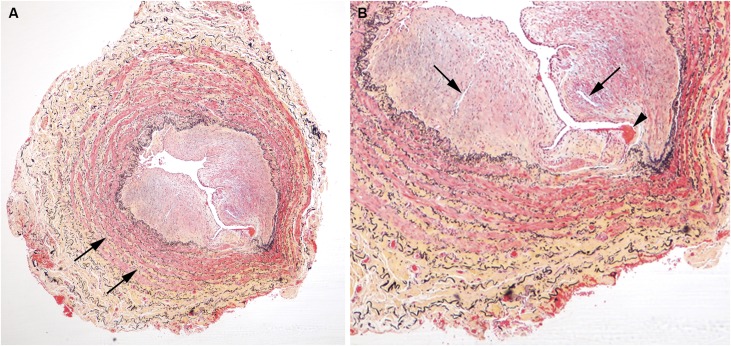
Figure 5.
Example of vein, stained with Movat pentachrome, showing highest degree of luminal occlusion by a neointima. (A) This vein shows accumulations of collagen (yellow-stained matrix, arrows) dispersed throughout the media. (B) High-power view of vein in (A) shows a small thrombus (arrowhead) at the neointimal surface. Several small vessels within a more cellular region of the neointima are present (arrows). The IEL (black stain) is disrupted in the portion of the vein near the thrombus.

