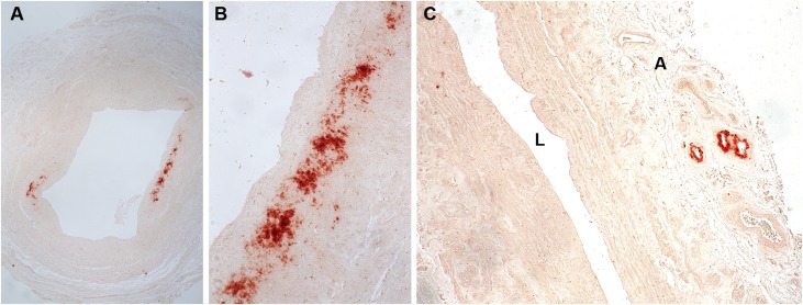
Figure 9.
Examples of calcification in vein detected by alizarin red S reagent. A lower power image (A) shows discrete areas of calcium accumulation (dark red). A higher power image of the calcium deposits in (A) is shown in (B). In (C), another vein sample had calcium deposits confined to neovessels in the adventitia (A). Calcification was typically detected in the medial and adventitial layers of veins with little accumulation of calcium within neointimas. A, adventitia; L, vein lumen.

