Figure 4.
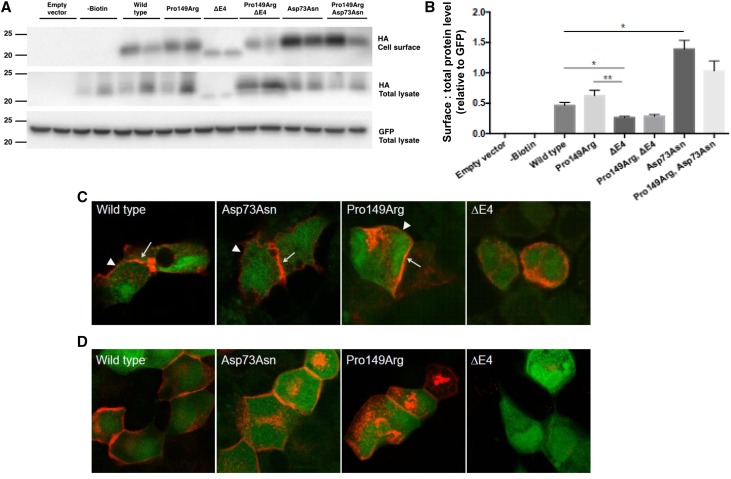
Variable response of Claudin-10b variants on cell surface expression and tight junction strand formation when expressed in HEK293 and MDCK-C7 cells. (A) Representative immunoblots of Claudin-10b variant–transfected HEK293 cell surface and total lysates probed for the hemagglutinin (HA) tag or green fluorescent protein (GFP) expressed with the Claudin-10b variant. (B) Densitometry of immunoreactive bands quantified from three independent experiments expressed relative to GFP as an indication of successful transfection. Statistical analysis was performed with an unpaired t test. *P≤0.05; **P≤0.01. (C) Immunohistochemistry of HEK293 cells lacking endogenous claudin expression and transiently transfected with Claudin-10b wild type, Asp73Asn, and Pro149Arg (red) enriched at cell-cell contacts (arrows) compared with cell membranes without contact to a transfected cell (arrowheads), indicating autonomous tight junction formation. In contrast, Claudin-10b ΔE4 was not localized within the cell membrane and thus, could not form tight junction strands. GFP is shown in green. (D) Immunohistochemistry of MDCK subclone C7 cells with endogenous claudin expression and tight junction formation transiently transfected with Claudin-10b variants, Claudin-10b wild type, Asp73Asn, and Pro149Arg (red) integrated in the cell membrane. Claudin-10b ΔE4 was only weakly expressed and did not localize to the cell membrane. GFP is shown in green.

