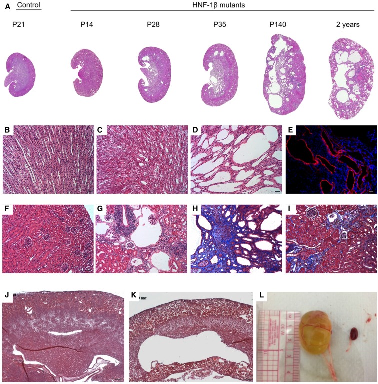
Figure 1.
Characterization of CD-specific HNF-1β mutant mice. (A) Hematoxylin and eosin (H&E) staining of kidney sections from control mice and HNF-1β mutant mice at the indicated ages. All images were acquired at the same magnification. (B and F) Higher magnification images of control kidneys at P14 showed normal histology of (B) the medulla and (F) the cortex. (C and D) Higher magnification images of HNF-1β mutant kidneys showed (C) a few dilated tubules at P14 and (D) significant cyst formation at P35. (E) Staining with an antibody against aquaporin-2 (red) showed that the cysts originated from the CD. Nuclei were counterstained with 4′6-diamidino-2-phenylindol (blue). (G) At 20 weeks of age (P140), the majority of HNF-1β mutant mice also exhibited glomerular cysts. (H and I) Trichrome staining of kidneys from mutant mice at 20 weeks revealed interstitial fibrosis (blue; H) surrounding medullary cysts and (I) in the renal cortex. (J and K) H&E staining showed that hydronephrosis was (K) present in some mutant mice at P7 and (J) absent in control littermates. (L) Hydronephrosis was more severe and frequent in mutant mice at later ages (P140). A control kidney is shown to the right. Scale bars, 50 μm in B–D and F–I; 20 μm in E; 100 μm in J–K.

