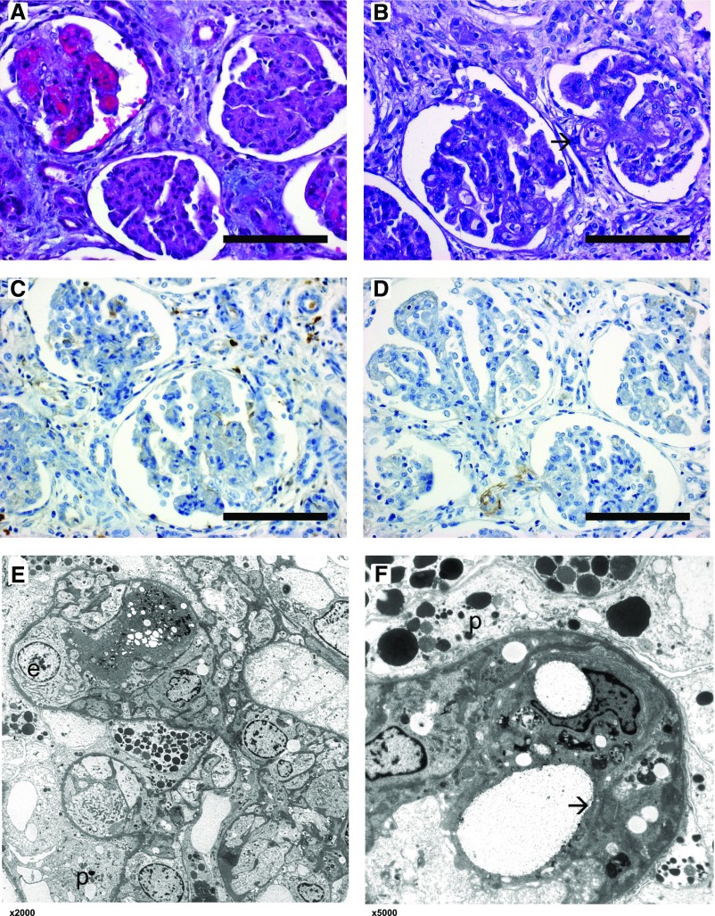
Figure 2.
Renal histology of patient 2.1 shows features of both MPGN and TMA. Light microscopical changes of affected glomeruli with lobulation of the glomerular tuft, thickening and double contours of glomerular basement membranes, collapse of the capillary tuft, and swelling of endothelial cells. One glomerulus shows active TMA (arrow). (A) Hematoxylin and eosin stain, original magnification, ×40; (B) periodic acid–Schiff stain, original magnification, ×40. (C and D) Immunohistochemistry using antibodies against IgG and C3c shows no specific glomerular staining. Original magnification, ×40. (E and F) Electron microscopy at various magnifications (×2000, ×5000) showing endothelial cell swelling (e) with subendothelial cleft formation (arrow) and capillary lumen obliteration. Glomerular basement membrane is intact. Podocytes (p) show enlarged cytoplasm and foot process effacement. No specific osmiophilic deposits and no fibrils.

