Figure 1.
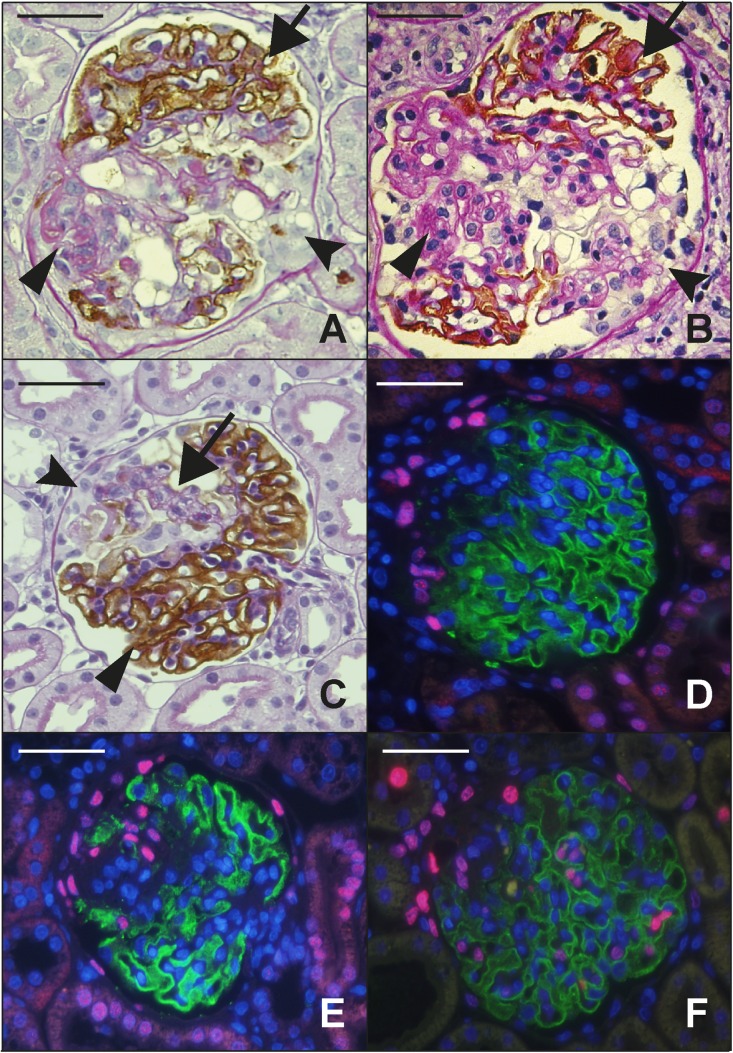
Structural features related to FSGS lesion development. (A) TG rat glomerulus showing a mature FSGS lesion at 7 months by Glepp1 peroxidase counter-stained with PAS. Podocytes are absent from the sclerotic area (arrowhead) in contrast to the remainder of the tuft which shows a normal distribution of brown peroxidase positive Glepp1-stained podocytes (arrow). An adhesion joining Bowman’s capsule to the glomerular tuft is also present (fluted arrowhead). (B) A mature human FSGS lesion that is identical to the rat FSGS lesion shown in (A). The arrows point to the same features as emphasized for (A). (C) Early segmental FSGS lesion present at 3 weeks after nephrectomy of the AA-4E-BP1 rat model. A naked glomerular capillary loop (arrow) is devoid of the brown-staining Glepp1-peroxidase product that is present in the remaining tuft area (arrowhead). PECs accumulate on Bowman’s capsule adjacent to the segmental lesion and some PECS appear to have invaded the tuft within the segmental lesion. (D and E) PECs, identified by their pink Pax8-positive nuclei. In (D), Pax8-positive cells have accumulated opposite an early developing FSGS lesion that is losing its podocytes (as indicated by loss of green Glepp1-stained cytoplasm). In (E), the Pax8-positive PECs are invading the glomerular tuft at a site of absent podocytes (as shown by absence of Glepp1 green fluorescence). Nuclei are shown by blue DAPI staining. (F) Ki67 immunofluorescence to identify dividing cells. In association with development of the early FSGS lesion as indicated by loss of Glepp1-positive green signal, Ki67-positive nuclei representing cycling cells (pink nuclei) are present on Bowman’s capsule (PECs), within the glomerular tuft (intrinsic glomerular cells), in the peri-glomerular and interstitial compartments, and in tubules. Double-labeling studies for WT1 and Ki67 showed no double-positive cells, indicating that cells identified as podocytes on the basis of anatomic location were not WT1-positive podocytes. Class II staining showed no increase in class II positivity in association with FSGS lesion formation. Scale bars, 100 μm.

