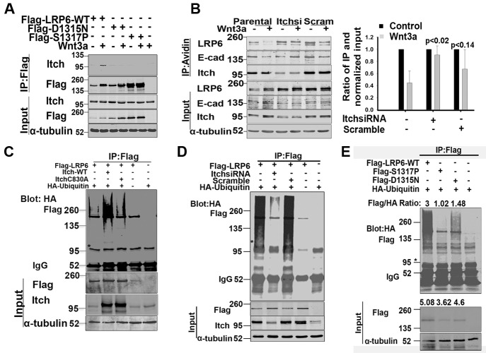
Figure 4. LRP6 ubiquitination by Itch E3 ligase.
(A). Itch interaction with LRP6. 293T cells transfected with LRP6 WT or LDLRR mutant were treated with Wnt3a for 1hr. Cells were then lysed using NP40 buffer and processed for IP with Flag antibody followed by immunoblotting. (B). Effect of Itch knock down on LRP6 internalization. 293T cells were transfected with either siRNA targeting Itch or control scramble siRNA. 48 hrs post-transfection, cells were labelled with cell impermeable biotin and then treated with Wnt3a for 1hr. Cells were processed for IP with avidin conjugated beads followed by immunoblotting as in Figure 3E. The bar graph was generated as in Fig.3D. Each bar presents data from one sample and error bars are SEM obtained from independent experiments run at similar conditions (n=3). The p values were calculated using unpaired two-tailed t-test comparing values of Wnt3a treated Itch and scramble siRNAs samples to Wnt3a treated parental sample. (C). Ligase active Itch ubiquitinates LRP6. 293T cells were co-transfected with flag tagged LRP6-WT and HA tagged ubiquitin and 24 hrs later trypsinized. Equal numbers of cells were replated and 24 hrs later transfected with Itch wild type or ligase dead ITCH C830A. At 40 hrs following the second transfection, cells were lysed in the presence of 1% SDS and processed for IP with Flag conjugated beads followed by immunoblotting. The same blot was probed sequentially with HA and flag using Licor secondary antibodies with either 700 or 800 spectra. *An unidentified band at 95kDa is seen. (D). Endogenous Itch ubiquitinates LRP6. 293T cells were co-transfected with HA tagged ubiquitin and flag tagged WT LRP6. Cells were trypsinized 24hrs post transfection, replated and transfected 24 hrs later with either Itch siRNA or scramble siRNA. 48hrs post second transfection, cells were processed as in Figure 4C. (E). Ubiquitination of LRP6-LDLRR mutants. 293T cells were transfected with HA tagged ubiquitin, and 24hrs post transfection were trypsinized and replated. 24hrs later, the cells were transfected with LRP6 WT or LDLRR mutant. Cells were processed for IP and immunoblotting as in Figure 4C. Flag/HA ratios are pixel values obtained from Flag IP normalized to HA from IP for each sample. In the WCL, the ratios are from Flag and tubulin. The blots shown are representative of independent experiments conducted for reproducibility (n≥2). Western blots were processed using Photoshop to adjust brightness/contrast and cropped to show all important bands.

