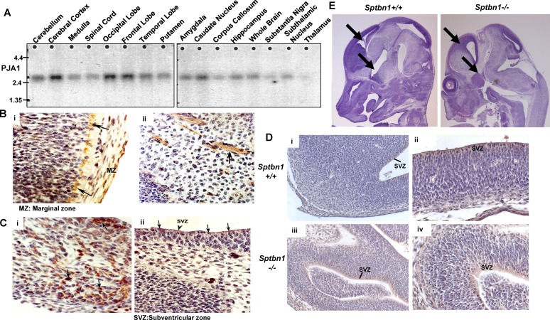
Figure 2. PJA1 expression in brain.
A. Northern blot analysis of PJA1 expression in human brain tissues. Cerebral cortex, occipital, frontal lobes and caudate nucleus demonstrate prominent mRNA for PJA1. B. Immunoperoxidase labeling of PJA1 in E12 mouse embryo forebrain marginal zone (MZ) (i) and mantle zone (ii). The left panel (i) shows Cajal-Retzius cells of the marginal layer labeled (arrows) while the right panel (ii) shows fibrillary label extending from cells in the mantle zone. C. Immunoperoxidase labeling of PJA1 in E12 mouse embryo midbrain (i) and cerebellum (ii). PJA1 is expressed in the SVZ cells and in developing midbrain nuclei (arrows). D. PJA1 expression compared in E12 forebrain of wild type (i-ii) and mutant Sptbn1−/− mice (iii, iv). Left panels are (i, iii) at 20X magnification; right panels (ii, iv) are at 40X magnification. PJA1 is expressed in SVZ cells and appears to be more widely distributed in embryonic brain of mutant Sptbn1−/− mice. E. Immuno-histochemical analysis of H & E staining of mouse embryos reveals the fore brain development defect in mutant Sptbn1−/− mice.

