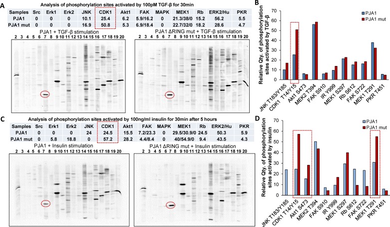
Figure 5. Elevated Cdk1 activity is associated with loss of PJA1.
A. Increased Cdk1 phosphorylation in cells transfected with PJA mutant. Protein phosphorylation visualized by ECL show increased phosphorylation activity in Cdk1 (Lane 7) in TGF-β treated cells transfected with PJA1 mutant (ΔRING mut) (right) compared to that in cells transfected with wild type PJA1 setting (left). The quantification results of bands were showed in the top table. B. Bar graph shows the quantification of the phosphorylation of each kinase and their respective sites as indicated in A. C. Increased phosphorylation activity in Cdk1 (Lane 7) in insulin treated cells transfected with PJA1 mutant (ΔRING mut) (right) compared to cells transfected with wild type PJA1 setting (left). The quantification results of bands were shown in the top table. D. Bar graph shows the quantification of the phosphorylation of each kinase and their respective sites as indicated in C.

