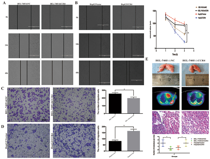
Figure 4.
CCR4 promotes HCC cells metastasis in vitro and in vivo. (A and B) Wound-healing assay shows a significant decrease or increase in healing rate of the scramble wound in BEL-7405/shCCR4 and HepG2/CCR4 respectively. (C) Silencing CCR4 in BEL-7405 cells could reduce the migrated cells through transwell assay. (D) Overexpress CCR4 in HepG2 cells could significantly increase the migrated cells through transwell assay. (E) Typical image of the effect on lung metastases of HCC cells via tail vein injection. The arrows in the upper panel indicate lung metastasis tumors. Representative images of 18F-FLT micro-PET/CT images of mice are shown at the middle panel, arrow indicates 18F-FLT uptake positivity in thoracic metastatic lesions. While the pathological image showed in the lower panel, arrow indicate metastatic tumors. *p < 0.05, **p < 0.03, ***p < 0.01. Data represent the mean ± SD and are representive of three independent experiments.

