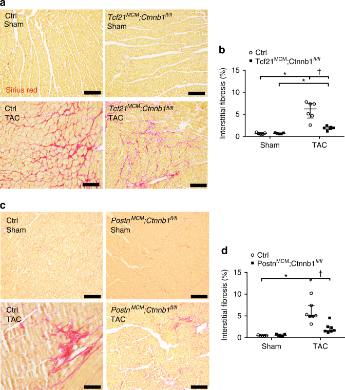
Fig. 4.
Loss of β-catenin in Tcf21 or Postn lineages leads to reduced interstitial cardiac fibrosis 8 weeks after TAC. a, c Heart sections were stained with Sirius Red to visualize fibrosis (red). Representative images of interstitial fibrosis are shown for Tcf21 MCM ;Ctnnb1 fl/fl (a) and Postn MCM ;Ctnnb1 fl/fl (c) with corresponding Cre-negative controls 8 weeks after sham or TAC operations. Scale bar = 100 μm. b, d The percent total area of interstitial fibrosis as indicated by Sirius Red staining was quantified. Data points are shown with median and interquartile ranges indicated. Statistical significance was determined by Kruskal–Wallis tests followed Mann–Whitney U tests for pairwise comparisons using Bonferonni adjustments to control for multiple testing. *P < 0.05 vs. Sham, † P < 0.05 vs. Control TAC. N = 5–7 mice per group

