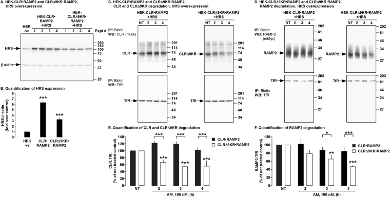
Figure 7.
Effect of HRS on the degradation of CLR, CLRΔ9KR and RAMP2. (A,B) Cell lysates from experimental cells were analyzed by Western blotting and levels of HRS and β-actin quantified. (C,D) Cell-surface biotinylated HEK-CLR•RAMP2 and HEK-CLRΔ9KR•RAMP2 cells expressing HRS were not treated (NT) or challenged with AM (100 nM, 2-4 h), biotinylated proteins immunoprecipitated (IP) and Western blots (WB) probed for CLR (mouse-HA, mHA), CLRΔ9KR (mHA), RAMP2 (mouse-Myc, mMyc) and transferrin receptor (TfR, loading control). In untreated HEK-CLR•RAMP2 and HEK-CLRΔ9KR•RAMP2 cells, CLR, CLRΔ9KR, RAMP2 and TfR were readily detected. AM (100 nM, 4 h) induced degradation of CLR, CLRΔ9KR and RAMP2 to different levels. (E,F) Quantification of degradation of CLR, CLRΔ9KR and RAMP2. n = 4, Data were examined using ANOVA and Student-Newman-Keuls post-hoc test, *p < 0.05, **p < 0.01, ***p < 0.001.

