
Figure 5. Increase in cell death upon loss of CYGB is O2 and NO-dependent in rat aortic vascular smooth muscle.
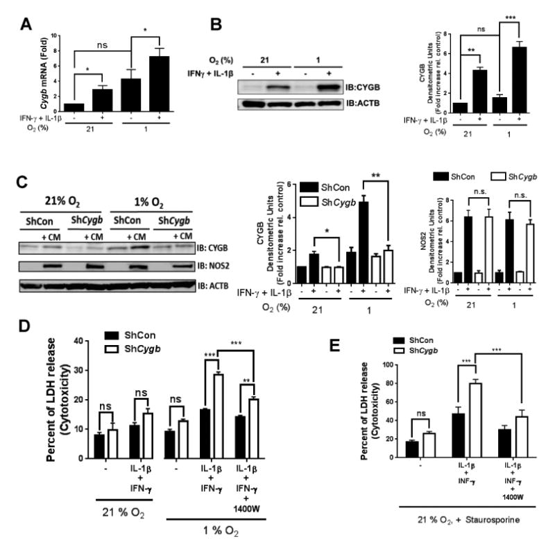
A) qRT-PCR results showing mRNA levels of CYGB in sub-cultured rat aortic vascular smooth muscle (RAVSM) cells. The cells were stimulated for 48 hours with interleukin-1β (IL-1β; 10 ng/ml) and interferon-γ (IFN-γ; 200 U/ml) at 21 or 1% O2. Mean ± SEM, n = 8. B) WB results showing protein levels of CYGB in sub-cultured RAVSM using the same conditions described in (A). Densitometric analysis is shown on the right panel. Mean ± SEM, n ≥ 6; for (A) and (B) *P <0.05, **P<0.01, and ***P<0.001, as determined by single or paired sample t-test with Bonferroni correction. C) Western blot results showing protein levels of CYGB and NOS2 in rat aortic VSM cells in the presence of a cytokine mix (CM, IL-1β + IFN-γ) with or without adenoviral silencing of CYGB.. Densitometric analysis is shown on the right panels. Mean ± SEM, n = 4;*P <0.05, **P<0.01, as determined by paired sample t-test with Bonferroni correction. D) Cells were stimulated with IL-1β and IFN-γ for 48 hours at 21 or 1% O2 with or without silencing of CYGB and cytotoxicity was determined by measuring LDH release. Mean ± SEM, n =3 E) cells were stimulated with IL-1β and IFN-γ for 48 hours at 21% O2 with or without silencing of CYGB and incubated with staurosporine for 24 hours before cytotoxicity was determined by measuring LDH release. Mean ± SEM, n =5. for (D) and (E) **P<0.01, and ***P<0.001, as determined by 2-way ANOVA.