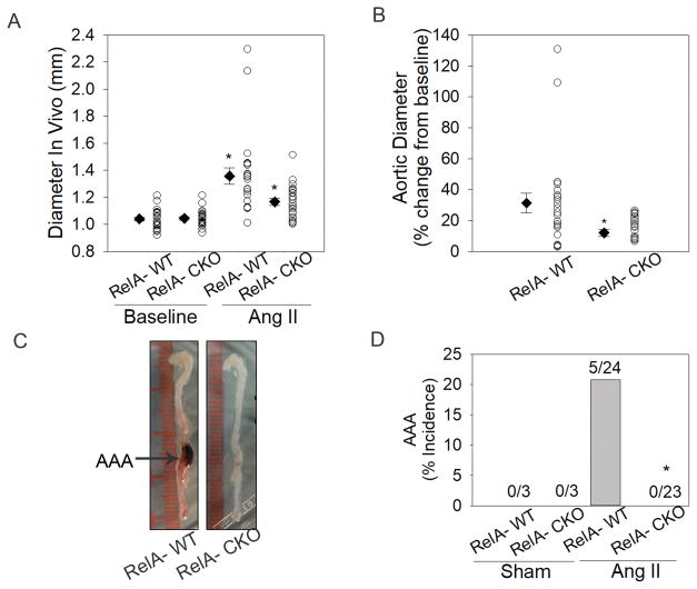
Figure 4. RelA depletion in aortic VSMCs and fibroblasts protects for AAA.
A, Abdominal aortic diameter measurements of RelA-WT (n=13) and RelA-CKO (n=14) mice were recorded at baseline and during day 6 of Ang II infusion using ultrasonogropahy. Circles represent measurement of individual mice and diamonds with error bars represent the mean ± SEM of the group. *P< 0.05 vs. RelA-WT at baseline; #P< 0.05 vs. RelA-WT with Ang II. B, Change in abdominal aortic diameter during Ang II infusion is presented as percent change from baseline. Circles represent individual mice and diamonds with error bars represent the mean ± SEM. *P< 0.05 vs. RelA-WT. C, Representative images of aortas from Ang II-infused mice. RelA-WT aortas developed AAA whereas RelA-CKO aortas remain protected from this disease. D, Percent incidence of AAA in RelA-WT and RelA-CKO infused with saline (sham) or Ang II for 7 days. Data are cumulative of three independent experiments. *P<0.05 vs. RelA-WT treated with Ang II, Fisher’s exact test.

