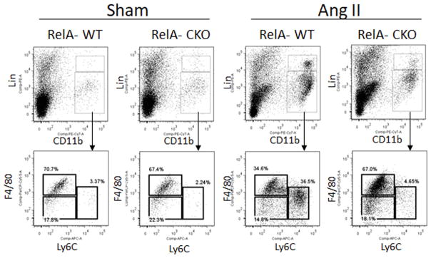
Figure 6. RelA deletion limits recruitment of inflammatory monocytes into the aortic wall.
Aortas from RelA-WT and RelA-CKO mice infused with saline (sham) or Ang II for 7 days were subjected to flow cytometric analysis for monocytes. Monocytes were identified as CD11bhi, Linlo, and F4/80lo. They were further subdivided based on their expression level of Ly6C into Ly6Chi and Ly6Clo. Macrophages were identified as having F4/80hi expression. Lin represents a combination of cell surface markers including B220, CD49b, CD90, NK1.1 and Ly6G. n=3–5 mice per group.

