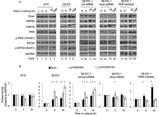
Figure 3. Elevated dicer protein levels observed during mild (39.5°C) hyperthermia-induced thermotolerance are linked to PKR and eIF2α phosphorylation in HeLa cells.

(A) Western blot analysis of dicer, phosphorylated and total PKR and eIF2α at 37°C and at 39.5°C in the presence of control or dicer siRNA. Mild hyperthermia treatments were initiated 48h post-transfection. TNF-alpha (10 ng/ml for 2h) was used as the positive control for phosphorylated PKR and eIF2α. A PKR inhibitor (250 nM) was added 1h before mild hyperthermia treatments were initiated. The lane ‘L’ indicates a commercial source of HeLa cell lysate that was used as a comparator to confirm that cells used in our experiments did not have detectable levels of active PKR in the absence of cell stress. PKR inhibitor was added to samples heated for 0h, 6h and 16h (i.e. lanes 17, 18 and 19). (B) Protein band intensities in (A) were first normalized to the GAPDH band intensities corresponding to that specific protein and further normalized to the 0h time point in the respective experimental group. Breaks in western blot images were created to juxtapose time points pertinent to the experimental question. The ratios of phosphorylated to total proteins are graphed. The representative western blot images for GAPDH correspond to the western blot image for dicer this figure. Values shown are mean ± SEM. n = 3, *p < 0.05.
