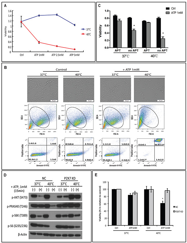
Figure 1. Hyperthermia potentiates ATP cytotoxicity in a P2X7-dependent manner.

(A-B) MCA38 wild type (WT) cells were treated with exogenous ATP at indicated concentrations and temperatures for 15 min. Cells treated with media served as control (Ctrl). Cell viability was determined using CCK-8 (A) and images of live cells were captured by Celigo (upper) and cell apoptosis/necrosis was evaluated by FACS (middle and bottom) (B). (C) Cells were left untreated or pre-treated with 2.5 μg/ml of APT102 (soluble ectonucleotidase CD39) for 30 min before exposed to ATP 1 mM for 30 min at indicated temperatures. Cell viability was measured 24 hr later. (D-E) MCA38 P2X7 KD (P2X7-deficient) and NC (negative control) cells were treated with ATP (1 mM for 15 min) at 37°C or 40°C. AKT/PRAS40/mTOR signaling pathway was analyzed by Western blotting (D) and cell viability was evaluated after 24 hr (E). *p < 0.05 as compared to control (one-way ANOVA, followed by Tukey pos-test). Bars, 50 μM.
