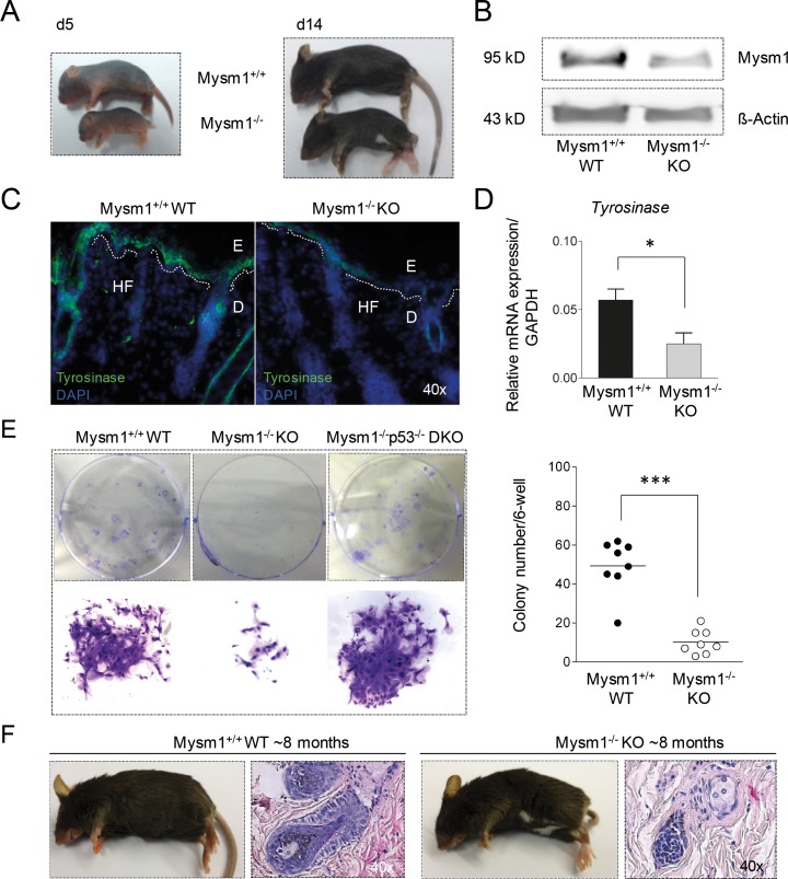
Figure 1. Pigmentation defect and altered melanocyte specification in Mysm1−/− mice.
(A) Postnatal development of visible skin pigmentation in Mysm1−/− (KO) in comparison with wild-type (WT) mice. The belly-spot-and-tail-phenotype manifested around 14 days after birth of Mysm1−/− mice. (B) Mysm1 protein expression in murine skin by Western Blot relative to Δ-Actin. Low residual Mysm1 expression in KO mice reflects hypomorphic Mysm1 alleles. (C) Tyrosinase expression in newborn Mysm1−/− skin compared with WT littermates (Tyrosinase green, DAPI-stained nuclei blue, dotted white lines separate E:epidermis and D:dermis, HF:hair follicle, n≥3, representative slides shown). (D) qPCR analysis of Tyrosinase mRNA expression in newborn Mysm1−/− and WT control mice relative to GAPDH. Bar graphs show mean results of 3 independent experiments with at least 3 mice of each genotype, standard deviations as indicated. (E) Reduced colony formation potential of precursor cells derived from skin of 8-week-old Mysm1−/− compared with WT and p53−/−Mysm1−/− DKO mice under melanocyte differentiation condition (n>4). (F) Monitoring of hair graying in adult 6- to 9-month-old Mysm1−/− and WT mice (representative photos shown, n>6 each genotype) and H/E stainings of corresponding skin samples (original magnification 40x).

