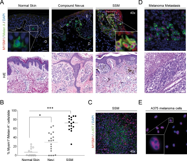
Figure 2. MYSM1 expression in normal human skin, nevi, primary melanoma, and melanoma metastases.
(A) Representative microphotographs of IF analyses of normal skin, dysplastic nevi from 6 patients, or SSM sections from 10 melanoma patients, and corresponding Hematoxylin&Eosin stainings from the same sections. The white dotted lines indicate the borders between epidermis (E) and dermis (D). MYSM1 protein expression was up-regulated in the nuclei of Melan A-positive cells in SSM and to a lesser extent in nevi compared with normal skin (MYSM1 red, Melan-A green, DAPI-stained nuclei in blue, original magnification as indicated). (B) Quantification of Melan-A+MYSM1+ double-positive cells in normal skin, melanocytic nevi (n=6) and SSM (n=10). (C) Co-localization of MYSM1 and c-KIT in selected SSM samples (MYSM1 red, c-KIT green, DAPI-stained nuclei blue). (D) MYSM1 protein expression in melanoma skin metastases identified by double-staining of MYSM1 and Melan-A (MYSM1 red, Melan-A green, DAPI-stained nuclei blue, representative IF, n=6). (E) Localization of MYSM1 protein in A375 melanoma cells in culture (MYSM1 red, DAPI-stained nuclei blue, representative photo, n>6).

