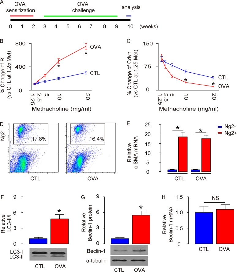
Figure 1. Enhanced ASM cell autophagy is detected in mouse lung after OVA treatment.
(A) Schematic of OVA model. Mice were first sensitized to alum-adsorbed OVA for 2 weeks, and then exposed to repeated airway provocation for another 7 weeks to establish airway hyper-sensitivity. (B) Dose-dependent responses in lung resistance (Rl) to methacholine. (C) Dose-dependent dynamic compliance (Cdyn) in response to methacholine. (D) Representative flow charts for purification of ASM cells from lung digests at the end of 7-weeks’ OVA challenge, based on Ng2 expression. (E) RT-qPCR for α-smooth muscle actin (α-SMA) in Ng2+ and Ng2- cells. (F) Western blotting for LC3 in Ng2+ and Ng2- cells. (G-H) Western blotting (G) and RT-qPCR (H) for Beclin-1 in Ng2+ and Ng2- cells. *p<0.05. NS: non-significant. N=10.

