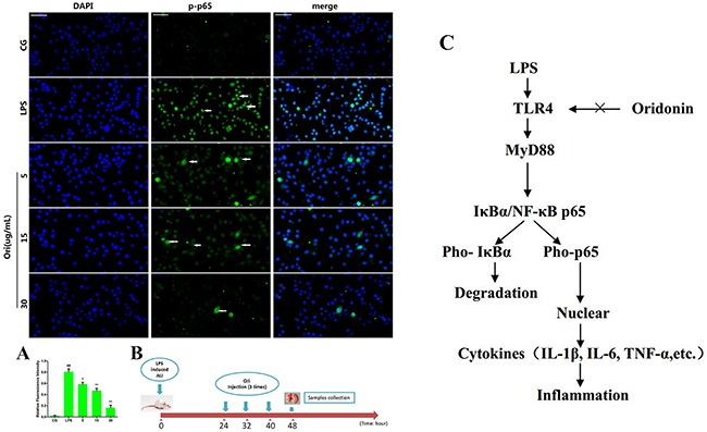
Figure 8.

(A) Translocation of the p65 subunit from the cytoplasm into the nucleus was evaluated by immunofluorescence. Blue spots represent cell nuclei, and green spots represent p-p65 staining; scale bar: 50 μm. The integrated option density (IOD) of DAPI was used as an internal control. All of the data represent the mean ± SEM (n=3).#p< 0.05, ##p< 0.01 versus CG; *p< 0.05, **p< 0.01 versus LPS. (B) Time axis of experimental animal treatment. (C) Schematic diagram of the signalling pathway related to the anti-inflammatory effects of Ori on LPS-induced ALI. LPS can induce NF-κB activation via TLR4-MyD88 signalling, IκBα acts as an inhibitor of NF-κB, Once the pathway is activated and IκBα is degraded, the NF-κB subunit p65 translocates from the cytoplasm to nucleus, which triggers the transcription of target genes, including TNF-α, IL-1β, and IL-6, and thus regulates inflammatory responses. However, Ori attenuates the release of pro-inflammatory cytokines by inhibiting TLR4/MyD88/NF-κB activation.
