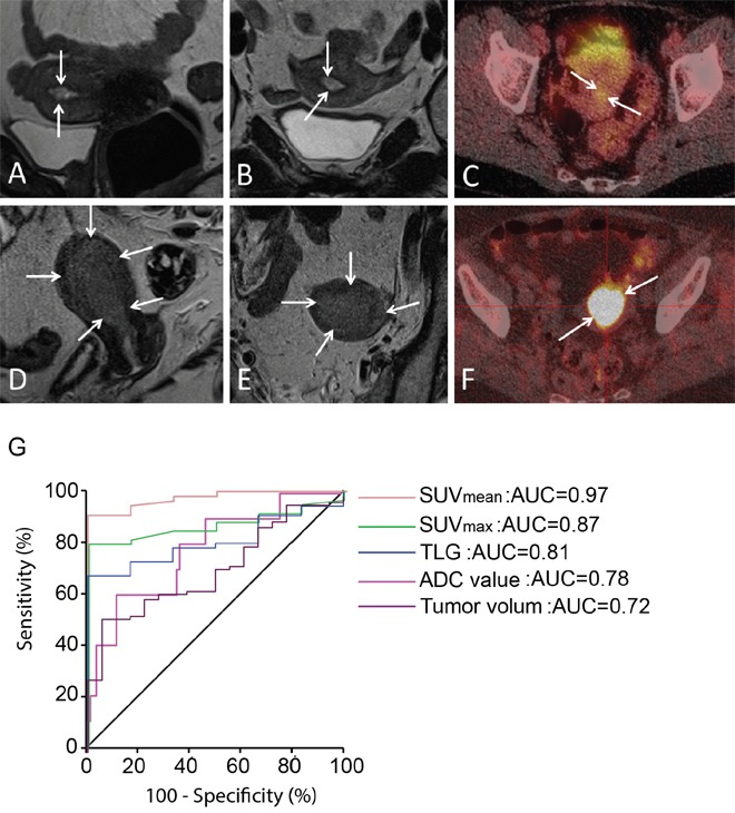
Figure 1. Preoperative imaging in premalignant and malignant endometrial lesions.

(A-B) Sagittal (A) and axial (B) T2-weighted MRI depicting a small uterine lesion (arrows) with volume of 0.5 cm3 in CAH patient. (C) FDG-PET/CT in the same patient demonstrates that the lesion (arrows) exhibits low FDG avidity (with SUVmean of 1.7 g/ml). (D-E) Sagittal (D) and axial (E) T2-weighted MRI depicting a large uterine tumor (arrows) with volume of 18.7 cm3 in a patient with EECG1. (F) FDG-PET/CT in the same patient shows that the lesion (arrows) exhibits high FDG avidity (with SUVmean of 11.1 g/ml). (G) ROC curves for the different imaging parameters (PET/CT: SUVmean, SUVmax and TLG. MRI: ADC value and tumor volume) for the discrimination of EECG1 from CAH shows that SUVmean yielded the best area under curve (AUC=0.97).
