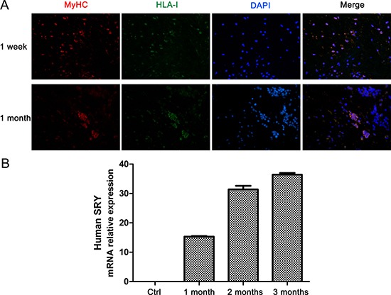
Figure 1. Grafted myoblasts survive and proliferate in heart tissue of minipig post-MI.

(A) Immunofluorescence staining of MyHC (red) and HLA-I (green) in heart sections from minipigs with MI at 1 week and 1 month after myoblast transplantation. Original magnification, ×400. (B) Q-PCR analysis of the expression of human Y chromosome in heart tissues of minipigs with MI followed by myoblast transplantation. Values are showed as the relative fold-change compared with control treatment groups (n = 6). Data are representative (A), or mean ± SEM (B) of 3 individual experiments.
