
FIGURE 10:
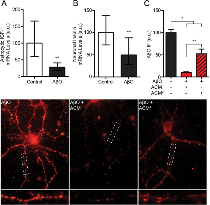
AβOs reduce insulin and IGF1 expression in astrocytes and neurons. (A) Treatment of cultured astrocytes with AβOs (500 nM) reduced IGF1 expression more than twofold (geometric mean = 28.0%; 95% CI = 19.1–40.8%) compared with control (geometric mean = 100%; 95% CI = 60.2–166%). (B) Treatment of cultured neurons with AβOs reduced insulin expression in neurons approximately twofold (geometric mean = 49.9%; 95% CI = 28.2–88.3%) compared with control (geometric mean = 100%; 95% CI = 72.3–138%). (C) Treatment of cultured astrocytes reduced the protective efficacy of conditioned media (based on images below). While conditioned media from untreated astrocytes (ACM) reduced neuronal AβO accumulation ∼90% (red bar), accumulation was down only ∼45% using media from astrocytes previously exposed to AβOs (AβO-ACM*; red and black striped bar). Geometric means and 95% CIs are plotted in A and B. Arithmetic means and standard errors are plotted in C. *, p < 0.05, Mann-Whitney; **, p < 0.01, Mann-Whitney.