FIGURE 1:
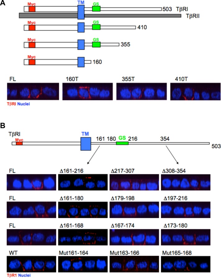
The basolateral localizing signal of the type I TGFβR is located at the juxtamembrane region between amino acids 161 and 164. (A) Top: Depiction of full-length (FL) TβRI and TβRII as well as three TβRI truncation mutants (TM, transmembrane domain; GS, glycine/serine rich domain; Myc, epitope tag). Bottom: Polarized MDCK cells were transiently transfected with the indicated FL or COOH-terminal truncated (T) TβRI constructs and visualized by confocal microscopy following staining for the extracellular Myc tag and secondarily with Cy3 (red) as described under Materials and Methods. Images are presented as perpendicular XZ cross-sectional images. Nuclei (blue) were stained with DAPI. (B) Top: Cartoon depicting location of tested regions relative to TM and GS domains. Bottom: Immunostaining of FL TβRI and indicated serial deletions (Δ) or alanine point mutations (Mut) in polarized MDCK cells. Row 1, deletions between amino acids 161 and 354. Row 2, deletions between amino acids 161 and 216. Row 3, deletions between amino acids 161 and 180. Row 4, wild type (WT) and alanine mutations between amino acids 161 and 168. Staining and visualization was as in A.

