FIGURE 3:
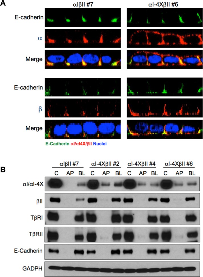
Chimeric TGFβRs confirm TβRI basolateral targeting motif. (A) Stable MDCK clones expressing either full-length (αIβII; clone #7) chimeric type I and type II TGFβRs or a full-length chimeric type II (βII) and a chimeric type I receptor containing alanine point mutations at residues V158, E161, E162, and D163 (αI-4X; clone #6) were transwell polarized and stained for either the extracellular GM-CSF α or β chain or E-cadherin as described (Anders and Leof, 1996; Murphy et al., 2004, 2007). Images are presented as perpendicular XZ cross-sectional images. Nuclei (blue) were stained with DAPI. (B) Biotinylation of endogenous TGFβRs and stably expressed chimeric TGFβRs in polarized MDCK cells. MDCK lines expressing wild-type βII either with αI or αIVEED/AAAA (αI-4X) were biotin labeled apically (AP) or basolaterally (BL) as described under Materials and Methods. Nonpolarized monolayer cultures were used to demonstrate total control labeling (C). Biotinylated proteins were extracted by streptavidin agarose beads and receptor specific antibodies were used for Western blotting. E-cadherin served as a basolateral marker, and GAPDH was used to confirm equal loading. Blots are representative of three separate experiments.

