FIGURE 4:
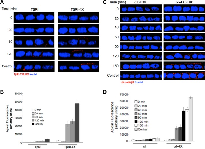
In the absence of the VEED motif, type I TGFβRs are directly targeted to the apical membrane. (A) Polarized MDCK cells transiently transfected with either native wild-type TβRI or TβRIVEED/AAAA (TβRI-4X) for 16 h were Golgi blocked at 20°C for 3 h in serum-free DMEM. Following washing with cold PBS the apical and basal chambers were then treated with a dilute (0.05%) trypsin/PBS solution for the last 30 min of the Golgi block to remove cell surface proteins (0 min). After a PBS wash, prewarmed fresh 10% FBS/DMEM was added, and the plates were returned to 37°C and stained for the Myc-tagged type I TGFβR at the indicated times after release. (B) Quantitation of apical receptor expression as observed in A presented as arbitrary fluorescence units ± SEM of 25 cells from three independent experiments. (C) αIβII and αIVEED/AAAAβII (αI-4XβII) MDCK cell lines were polarized on 12-mm Transwell plates. Subsequent to Golgi block and trypsinization as described in A, newly expressed chimeric type I TGFβRs were visualized by immunofluorescence from 20 to 150 min after release. Cells were stained for TβRI using primary antibody to the external GM-CSF α chain and secondarily stained by Cy3 (red). Images are presented as perpendicular XZ cross-sectional images. Nuclei (blue) were stained with DAPI. (D) Quantitation as performed in B of 30 cells from three independent experiments.

