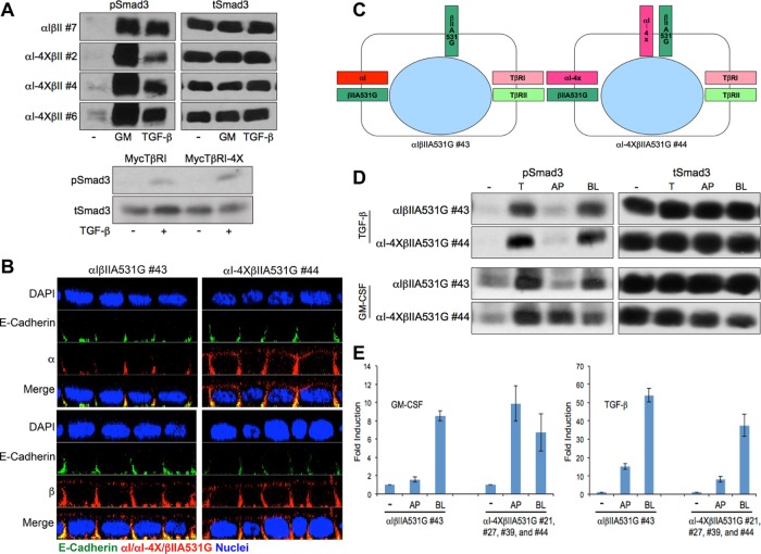
FIGURE 7:
Apical mislocalized TGFβRs are signaling competent. (A) Type I TGFβR basolateral targeting motif has no effect on TGF-β-induced Smad3 signaling. Top: Chimeric receptor expressing MDCK lines expressing wild-type (αIβII #7) type I and type II receptors or a wild-type type II receptor and type I receptor mutated in the VEED basolateral targeting domain (αI-4XβII #2, #4, and #6) were treated in the absence (-) or presence of GM-CSF (GM, 100 ng/ml) or TGF-β (10 ng/ml) for 1 h before being processed for Western analysis using phospho (p) or total (t) Smad3 sera. Bottom: R1B Mv1Lu cells (do not express native TβRI; Boyd and Massagué, 1989) were transiently transfected with either native wild-type TβRI or TβRI mutated in the VEED domain (TβRI-4X) directing basolateral receptor delivery. Phospho and total Smad3 was determined following 1 h stimulation ± 10 ng/ml TGF-β. (B) MDCK cells stably expressing either a wild-type chimeric type I receptor (αI) and a type II receptor (Murphy et al., 2007) mutated such that it mislocalizes to the apical membrane (αIβIIA531G #43) or chimeric type I and type II receptors that both undergo apical trafficking (αI-4XβIIA531G #44) were polarized on transwell inserts and stained for the indicated proteins. Images are presented as perpendicular XZ confocal cross-sections, and nuclei were stained with DAPI. (C) Cartoon depicting the locale of native and chimeric TGFβRs based on the chimeric receptors immunostaining data from B. (D) Chimeric clones from B were cultured in six-well transwells for 72 h. Cells were serum starved with 0.1% FBS/DMEM for 16 h and then either left untreated (-) or stimulated with TGF-β (10 ng/ml) or GM-CSF (100 ng/ml) from apical (AP), basolateral (BL), or both sides (T) at 37°C for 1 h. Equivalent protein was processed by Western blotting for phospho (p) or total (t) Smad3. Blots for A and D are representative of three separate experiments. (E) Polarized MDCK clones from D as well as three additional clones (e.g., #21, #27, and #39) expressing apically targeting chimeric TβRI and TβRII were treated as in D for 3 h with either GM-CSF (100 ng/ml; left) or TGF-β (10 ng/ml; right) and processed by RT-PCR for expression of PAI-1. Data reflect mean ± SEM from three biological replicates for control αIβIIA531G (#43) and pooled replicates for each (n = 8) of the αI-4XβIIA531G clones (#21, #27, #39, and #44).

