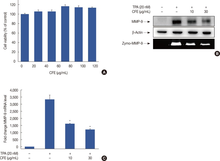
Figure 1. CFE inhibits TPA-induced MMP-9 expression in MCF-7 cells. (A) To assess the cytotoxicity of CFE, cells were treated with various concentrations of CFE for 24 hours. An EZ-cytox enhanced cell viability assay kit was used to detect the. (B) CFE inhibits TPA-induced MMP-9 expression in MCF-7 cells. MCF-7 cells grown in monolayer culture were treated with the indicated CFE concentrations in the presence of TPA for 24 hours. Cell lysates were analyzed by Western blot with anti-MMP-9 antibody and β-actin as a loading control. Conditioned medium was prepared and used for gelatin zymography (Zymo) to assess the effect of CFE on MMP-9 activity in MCF-7 cells. Cells were pretreated with CF for 1 hour and then stimulated with TPA for 24 hours. (C) MMP-9 mRNA levels were analyzed by RT-qPCR, and GAPDH was used as an internal control. Values are shown as mean±SEM of three independent experiments.
CFE=Crotonis fructus extract; TPA=12-O-tetradecanoylphorbol-13-acetate; MMP-9=matrix metalloproteinase-9; Zymo=zymography; RT-qPCR=real-time quantitative polymerase chain reaction; GAPDH=glyceraldehyde 3-phosphate dehydrogenase; SEM=standard error of the mean.
*p<0.01 vs. TPA.

