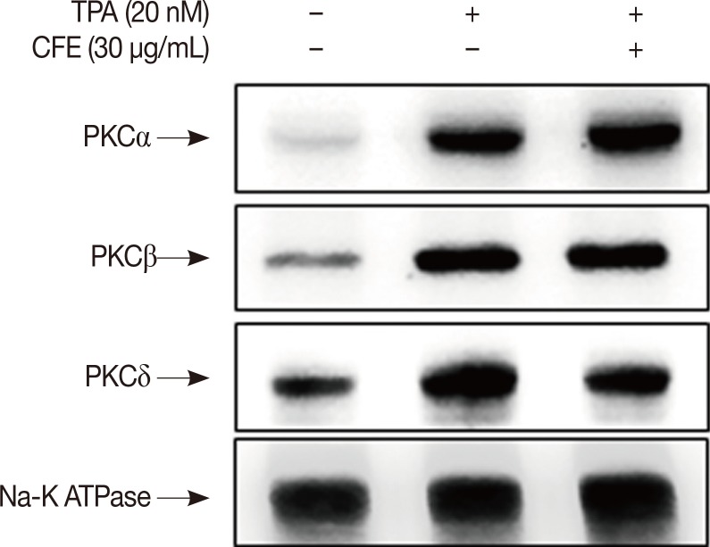
Figure 2. CFE inhibits TPA-induced activation of PKCδ. MCF-7 cells were pretreated with CFE for 1 hour and then with TPA for 1 hour. Western blot analysis was performed to detect the levels of PKCα, PKCβ, PKCδ, and Na-K ATPase as a loading control in the membrane fractions.
CFE=Crotonis fructus extract; TPA=12-O-tetradecanoylphorbol-13-acetate; PKC=protein kinase C.

