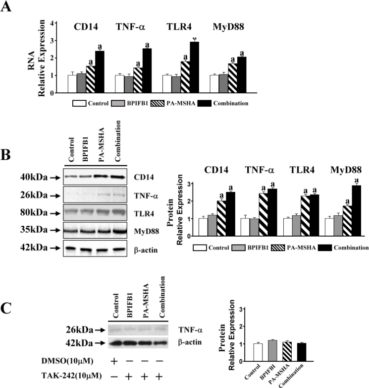
Figure 2.
CD14/TLR4/MyD88 pathway contributed to the PA-MSHA-BPIFB1 signaling transduction. A. 5×10⁵/ml differentiated THP-1 cells were treated with 2×10⁷/ml of PA-MSHA and 1 mg/ml BPIFB1 for 24 hours. RNA was extracted and first-strand cDNA was synthesized using SuperScript First-strand Synthesis system. The cDNA was used as a template in real-time PCR reactions to analyze the expressions of CD14, TNF-α, TLR-4, and MyD88. B. The protein expressions of CD14, TNF-α, TLR-4, and MyD88 were detected by Western blot. β-actin protein was used as the loading control. Protein bands were detected using SuperSignal West Pico Chemiluminescent Substrate; the intensity of the bands was analyzed by ImageJ to show the expression levels of proteins quantitatively. C. Differentiated THP-1 cells were co-incubated with BPIFB1, PA-MSHA and TLR-4 specific kinase inhibitors (TAK-242, 10 µM) for 24 hours. 10 µM DMSO was added into the control group. The cell lysates were used to determine TNF-α production by western blot. Data representative results derived from a minimum of 3 independent experiments. a: p<0.05 compared with none PA-MSHA treatment.

