Figure 3.
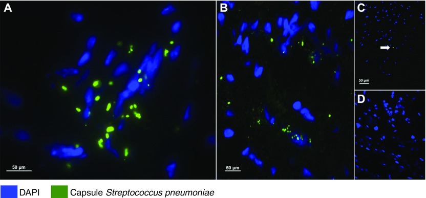
Streptococcus pneumoniae translocates into the heart during pneumonia. Representative images from (A and C) left ventricular and (B) interventricular septal sections from the six nonhuman primates (NHPs) with pneumococcal pneumonia and (D) three uninfected control animals. S. pneumoniae was visualized using immunofluorescent staining with antiserum against serotype 4 capsular polysaccharide (green) and 4′,6-diamidino-2-phenylindole (DAPI; blue) to reveal nucleated tissue cells. (A and B) Bacterial aggregates with diplococci morphology are seen within the left ventricles and septa of NHPs with acute pneumonia. (A) Bacterial cluster conformation indicates active replication. (C) Single-capsule debris (marked with the white arrow) were scattered throughout the left ventricles and in the interventricular septa of convalescent NHPs. (D) Pneumococcal capsules were not identified in the hearts of uninfected control NHPs.

