. 2017 Jun 14;8(8):5705–5712. doi: 10.1039/c7sc01380a

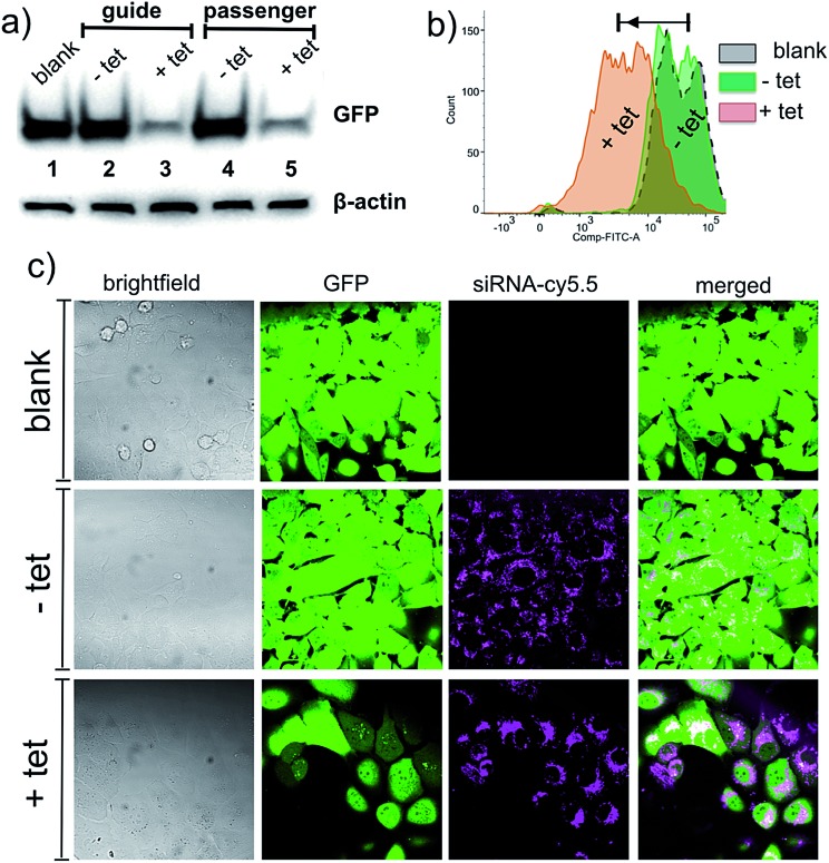
Fig. 2. Western blot analysis, flow cytometry and fluorescence microscopy measurements showing knockdown of GFP in MDA-MB-231-GFP cells. (a) Western blotting analysis of GFP levels, β-actin was used as a loading control; immobilization of siRNA on NP surface either through guide or passenger strand shuts down the activity of siRNA (lanes 2 and 4 vs. 1) whereas the addition of the tetrazine 2 activates the siRNA resulting in a remarkable decrease in the expression of GFP (lanes 3 and 5). (b) Flow cytometry analysis shows a decrease in the fluorescence of GFP signal upon addition of the tetrazine 2 (c) confocal fluorescence microscopy demonstrates that the siRNA (cy5.5) is taken up by cells and the loss in the GFP signal is observed with the addition of the tetrazine 2. The siRNAs tend to diffuse in the cell upon addition of the tetrazine 2, suggestive of release from the NP-TCO-siRNA.

Fig. 2