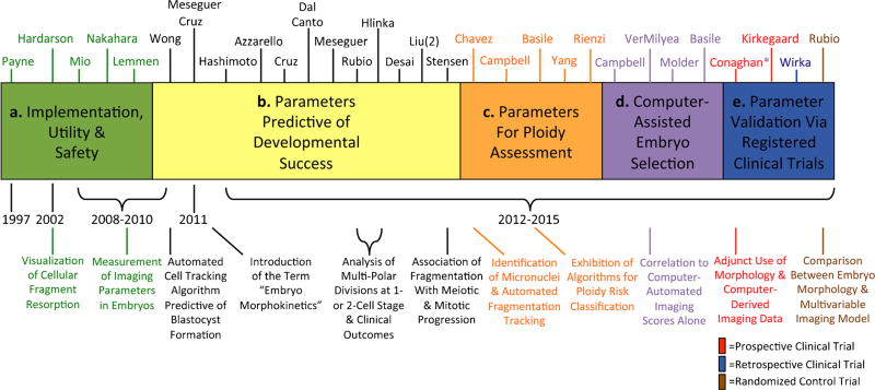
Fig. 2.
Evolutionary timeline of time-lapse imaging for embryo assessment. A timeline from 1997 to the present day showing the various “eras” of time-lapse image analysis with relevant references above and some of the major milestones below in the evaluation of embryo viability. The first time-lapse publications a examined the implementation, utility, and safety of this technology to monitor embryo development. Subsequently, studies began identifying cellular parameters predictive of success in the blastocyst stage b, followed by the identification of parameters predictive of embryo ploidy status c. More recent reports d have focused on the development of computer-assisted algorithms for automatic embryo assessment and classification. Other studies e have clinically applied and validated these parameters or newly identified parameters in either retrospective or prospective registered clinical trials. The first randomized control trial evaluating time-lapse image analysis versus traditional IVF assessment was recently published, with others likely to follow

