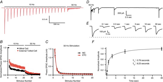
Figure 4. Single pool of slowly recycling vesicles.

A, representative external tufted cell recording showing 10 Hz stimulation followed by 50 Hz stimulation. B, group data showing immediate depression following 10 Hz stimulation, suggesting a single pool of synaptic vesicles in both mitral cells and external tufted cells. C, in both cell types, the phasic EPSC amplitude plotted as a function of stimulus number is fit by a single exponential decay. D and E, recovery of phasic EPSC amplitude following 50 Hz stimulation suggests that vesicle replenishment is slow. F, recovery time course (pooled data from both mitral and external tufted cells) is best fit by a double exponential. [Color figure can be viewed at wileyonlinelibrary.com]
