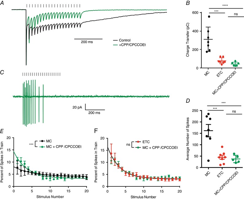
Figure 6. Blocking the slow current converts mitral cell responses into external tufted cell responses.

A, peak scaled comparison of the whole cell voltage clamp recordings from mitral cells in control and 10 μm CPP/20 μm CPCCOEt in response to 50 Hz ORN stimulation. As expected, CPP/CPCCOEt blocked a significant portion of the slow envelope current. B, comparison of the total charge transfer in mitral cells, external tufted cells and mitral cells with CPP/CPCCOEt shows that blocking the NMDA/mGluR1 receptor‐dependent current significantly reduces the total charge transfer to levels comparable to external tufted cells. C, cell‐attached recording from mitral cell in response to 50 Hz ORN stimulation shows transient spiking profile of mitral cells when NMDA and mGluR1 receptors are blocked. D, the total number of action potentials produced in mitral cells with NMDA and mGluR1 receptors are similar to external tufted cell responses. E, comparison of the temporal profile of mitral cell spiking in control and with CPP/CPCCOEt. Block of NMDA and mGluR1 receptors reveals transient response profile of mitral cells. F, with NMDA and mGluR1 receptors blocked, the temporal profile of mitral cell spiking is not significantly different from the responses of external tufted cells. [Color figure can be viewed at wileyonlinelibrary.com]
