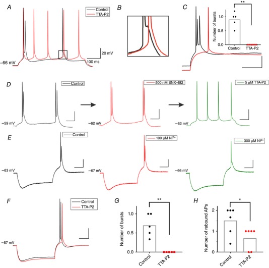
Figure 2. Pharmacological characterization of burst‐firing pattern in the rat subiculum.

A, original traces from a representative bursting neuron portraying the change in the firing pattern (from burst to regular‐spiking) as a response to a depolarizing 20 pA current injection, before (black) and after the addition of 10 μm TTA‐P2 (red). B, zoomed in trace from A, showing the elimination of ADP hump due to the application of TTA‐P2. C, original trace of rebound bursting (black) which was eliminated by 10 μm TTA‐P2 (red), and instead displaying a single rebound spike after 200 pA hyperpolarizing current injection. The inset graph represents the average number of rebound bursts before and after TTA‐P2 (* P < 0.05, ** P < 0.01, two‐tailed paired t test). D, original traces from a representative bursting neuron (Control; black trace) depicting the lack of effect of SNX‐482 (500 nm) on the burst‐firing pattern as a response to a depolarizing 30 pA current injection (red trace). When the low concentration of TTA‐P2 (5 μm) was added to the bath, the same neuron fired a regular train of action potentials (green trace). E, original traces of hyperpolarization‐induced bursting before (Control; black trace) and after 100 μm Ni2+ (red) that failed to attenuate bursting, followed by the same neuron with 300 μm Ni2+ (green), which successfully eliminated bursting, but without fully removing ADP. F, representative traces from a current‐clamp experiment performed at 33°C showing rebound burst firing in a rat neuron in control conditions (black trace) and after application of 5 μm TTA‐P2 (red trace). G, TTA‐P2 abolished rebound bursts in the experiment performed at 33°C. H, TTA‐P2 decreased the number of rebound APs in the experiments at 33°C.
